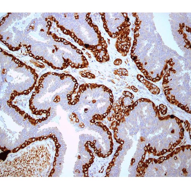
Immunohistochemistry is a technique used to determine the presence and level of specific cellular proteins. IHC measures protein expression using specially labeled antibodies that can bind to the proteins of interest. The antibody is mixed with the cellular components of the tumor. After a set amount of time, the mixture is rinsed and only those antibodies attached to their protein targets will remain. The presence of the antibodies can be detected by viewing the sample under a microscope because areas containing bound antibodies will appear a different color than areas lacking antibodies. Samples with more protein will bind more antibody and therefore appear darker. This allows the test to reveal not only whether a protein is present but also the relative amount of the protein. Test results are based on the strength of the staining and the percent of cells stained.1, 2, 3
IHC is a commonly used technique because it is inexpensive to perform, doesn't require special equipment, and generally quite accurate.2, 1, 3
As an example, three proteins of particular interest for breast cancer are HER2, the estrogen receptor (ER) and the progesterone receptor (PR).
HER2 is a growth factor receptor located on the surface of breast cells. About 30% of breast cancer patients have tumors that express a very large amount of HER2. The anti-cancer drug Herceptin® is targeted at this protein making accurate determination of the presence or absence of this protein in a tumor an important step in determining the appropriate treatment.
Learn more about Herceptin®
Learn More About HER2
The ER protein is another growth factor receptor. The ER protein binds to the female sex hormone estrogen and plays a major role in stimulating cell division in breast cells. Because drugs that interfere with estrogen signaling are an important tool in treating breast cancer, accurate determination of ER levels is important to the design of treatment plans.
Learn more about hormonal treatments for breast cancer
The PR protein is the receptor for the female sex hormone progesterone. While there are no targeted therapies directed against the PR protein, the presence or absence of the receptor in cancer cells is a factor in determining the prognosis of the disease. Learn More About ER and PR

Immunohistochemistry (IHC) is a method of detecting proteins in/on cells. Proteins that bind to the target are added to the cells and then colored proteins are added. If the target is present, the color will be detected with a microscope or other machine.
A Closer Look at Immunohistochemistry Results
Scoring system and assessment for HER2 status
- Scoring (from the makers of HercepTest™)
- (0) - one or little staining in < 10% of cells
- (1+) - faint, partial staining in > 10% of cells
- (2+) - weak to moderate, complete staining in > 10% of cells
- (3+) - strong, complete membrane staining in > 10% of cells
- Assessment of staining (according to the NCCN)
- Samples with 3+ (Not 2+/3+ borderline) are eligible for Herceptin™
- Samples with 2+ result should be retested using FISH
- Samples with 0/1+ result are HER2 negative
Scoring system and assessment for( )ER, PR status
- J-Score Method Scoring
- (0) - no stained cells
- (1+) - stained cells ≤ 1%
- (2+) - 1% < stained cells < 10%
- (3+) - stained cells ≥ 10%
- Assessment of staining
- Negative - Score of 0
- Indeterminate - Score of 1 or 2
- Positive - Score of 3
- Allred Method Staining
- (0) - no stained cells
- (1) - stained cells <1/100
- (2) - 1/100 ≤ stained cells < 1/10
- (3) - 1/10 ≤ stained cells < 1/3
- (4) - stained cells = 1/3 < 2/3
- (5) - stained cells > 2/3
- Intensity
- 0 = none
- 1 = weak
- 2 = intermediate
- 3 = strong
- Scoring
- To obtain the total score add staining score and intensity score. Any score between 0-2 is considered( )ER or PR negative, any score above 2 in considered( )ER or PR positive.
- 1ab Z Theodosiou, IN Kasampalidis, G Livanos, M Zervakis, I Pitas, K Lyroudia. Automated analysis of FISH and immunohistochemistry images: a review. Cytometry Part A. 2007. 71;7:439-50. [PUBMED]
- 2ab Zafrani B, Aubriot MH, Mouret E, et al. High sensitivity and specificity of immunohistochemistry for the detection of hormone receptors in breast carcinoma: comparison with biochemical determination in a prospective study of 793 cases. Histopathology. 2000; 37(6):536-45. [PUBMED]
- 3ab M Bilous, M Dowsett, et al. Current Perspectives on HER2 Testing: A Review of National Testing Guidelines. Modern Pathology. 2003; 16(2):173-182. [PUBMED]