Below is a list of the information included on this page:
History
The CT scan was first used clinically in the 1960's, but it wasn't until the late 1970's to early 1980's that medical use of CT was widespread. In 1979 two scientists, Godfrey Hounsfield and Allan Cormack, shared the Nobel Prize for their work developing the CT scanner. CT is also called Computerized Axial Tomography or CAT.1
How CT works
A CT scanner uses x-rays in the same way as a conventional x-ray but instead of taking one image a CT scanner takes multiple images, or slices. A computer program gathers all the images and compiles them to create a three dimensional image of the internal structures being examined. Advances have improved the sensitivity and usefulness of CT scanners since they were developed.1 2
CT Equipment
What instruments are used?
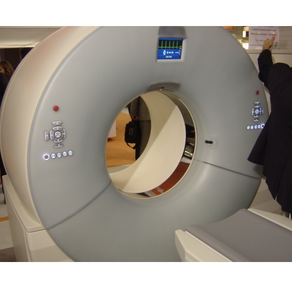
A CT scanner is made up of 4 main components, the gantry (frame) houses the X-ray source and detectors and has a large opening in the middle (patient port), the subject table which moves in and out of the patient port, the x-ray source and detectors, and a computer system that gathers information from the detectors and produces an image from the information.1
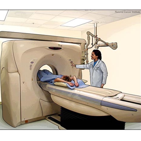
Image courtesy of the NCI
The CT Exam
Scheduling
When you are scheduling your exam be sure to mention if you:
- are breast feeding
- are pregnant or think you may be pregnant
- have a fear of enclosed spaces (claustrophobia)
- have metal implants of any kind
- have any kidney problems
- have any heart problems
- have had an adverse reaction to contrast dye
Day of the Exam
- Take your medications normally
- Depending on the type of exam, you may be asked to follow a certain diet
- Wear something comfortable, you may be asked to put on a gown
- Do not wear any jewelry; watches, chains, rings, piercings, etc.
During the Exam
The exact procedure used for a CT scan will depend on what area of the body is being scanned. You will need to remove anything metal from your body and may be asked to put on a gown. A CT technician will take you to the CT room where you will lay on a table connected to the CT scanner. You will be positioned on the table and the technician will start the scan. As the exam progresses you will slowly move through the scanner. The machine may make noise, this is normal; it is the x-ray unit and the detectors spinning around you. As the x-ray/detector unit spins around you it is capturing images, as many as 192 per second. The scan can last from 10 minutes to up to an hour, if you have trouble staying still or are claustrophobic you may need to be sedated.
In some cases you will be given a contrast material, this is necessary to obtain the best possible image. The contrast material will be given orally or intravenously.
After the Exam
You should have no side effects from the exam and should be able to resume your normal daily routine. If you needed a sedative you should arrange for a ride home from the imaging facility. If you had an exam involving contrast media you should drink plenty of water to get rid of any remaining contrast media in your body.
Contrast Media For CT Exams
What is it?
The contrast agents used in CT are usually an iodinated molecule. Iodine is very effective at absorbing radiation, this is why it is used in CT scans. Areas where the contrast media collects will produce a light image; this makes possibly dangerous lesions easier to see. Once injected/ingested, the contrast media is distributed throughout the body by the blood vessels and spreads into extracellular space. Only 1-2% of it actually enters the cells and none of it is metabolized, it is excreted unchanged through normal kidney processing.3

Image courtesy of the NCI
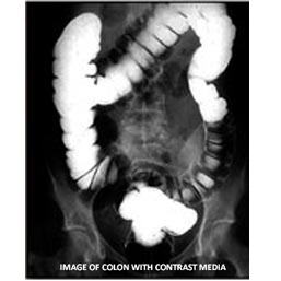
Image courtesy of the NCI
Procedure involving contrast agent
If your CT exam involves contrast media, you will need to follow some special procedures. You may be asked to follow a certain diet, which may include fasting. The contrast material can be given orally, intravenously, or both. If it is given orally you may be asked to drink it up to 24 hours before the exam and again shortly before the exam, so it is important to be on time for your exam. If the contrast media is given intravenously, a nurse will put an IV in your arm or hand before the exam. An IV injection of contrast material may make you feel flushed, warm, or nauseous and you may notice a metallic taste in your mouth. These are not serious issues and should subside. Serious reactions from the dye can occur, so tell the nurse about any other problems/feelings.3
CT Imaging Results
CT vs Conventional X-ray
Conventional x-ray lacks the ability to provide detailed information about internal structures. A major limitation of conventional x-rays is that the output is a 2-dimensional image of 3-dimensional structures. Additionally, many different body tissues have similar abilities to absorb x-rays. X-ray images are not able to detect differences between these tissues. X-ray is not an exact science and a large portion of the x-rays emitted are scattered (bounced away) from the patient, producing a less than complete image. CT resolves these problems; it can provide a 3-dimensional image of an internal structure, it can detect differences in tissue density, and the x-rays can be focused directly on specific areas, producing a clear image.1

Image courtesy of the NCI
Advances in CT
Spiral/Helical Computed Tomography (SCT)
A newer version of CT, Spiral CT, is capable of producing images more rapidly than conventional CT. The patient lies on a table that moves through a large opening in the machine. The x-ray source and detectors spin around the patient. In most CT scanners the source/detector unit rotates around the patient in less than 1 second, topping out at 330ms. Each rotation of the unit produces anywhere from 16 to 64 slices depending on the machine. These slices are compiled by a computer and combined to create a high resolution 3 dimensional image. Because each rotation can produce 64 slices and takes less than 1 second, the total time needed for a complete CT scan is generally less than 1 minute.4
An X-ray shoots beams at the targeted body area. Those parts that block the beam show up as light on the image (radiograph).

A CT machine produces a series of X-ray images - like slices of a tomato. The final image is a 3D model of the body region of interest (or a tomato in the animation above.
CT Cancer Risks
Computed axial tomography (CT scans or CAT scans) use X rays to create images of body tissues. X rays are a form of high energy radiation and are themselves, able to cause cancer. The doses of X rays used in CT scans are as low as possible, but they do present some risk of doing harm. This appears to be especially true in children. A 2012 study demonstrated that exposure to X rays via CT scans lead to an increased risk of leukemia and brain cancer in young patients. The overall number of people affected would not be expected to be large due to the rarity of the cancers that were caused.5
Learn more about radiation and cancer.
- 1 a b c d RA Robb. X-ray computed tomography: from basic principles to applications. Annual Review of Biophysics and Bioengineering. 1982;11:177-201. [PUBMED]
- 2Grignon B, Mainard L, Delion M, Hodez C, Oldrini G. Recent advances in medical imaging: anatomical and clinical applications. Surg Radiol Anat. 2012 May 29. [Epub ahead of print] [PUBMED]
- 3 a b MA Bettmann. Contrast media: safety, viscosity, and volume. European Radiology. 2005; 15 Suppl 4:D62-4. [PUBMED]
- 4WA Kalednder. X-ray computed tomography. Physics in Medicine and Biology. 2006; 51: R29-R43. [PUBMED]
- 5Pearce MS, Salotti JA, Little MP, McHugh K, Lee C, Kim KP, Howe NL, Ronckers CM, Rajaraman P, Craft AW, Parker L, Berrington de González A. Radiation exposure from CT scans in childhood and subsequent risk of leukaemia and brain tumours: a retrospective cohort study. Lancet. 2012 Jun 7. [Epub ahead of print] [PUBMED]